| art theory 101~SCoDNA: The Structure & Chemistryof DNA (axial view) |
|---|
from: PIN: Primes to DNA
© 2001, Reginald Brooks. All rights reserved.
- INTRODUCTION
- STRUCTURE
- CHEMISTRY
- CONCLUSION
[ As noted above, the main section of the Number Patterns in DNA has been given its own title, GoDNA: The Geometry of DNA , and a separate section to go to view this work. This section, has also been extracted from Patterns In Number (PIN), reworked and adapted for the web as: SCoDNA: The Structure & Chemistry of DNA (axial view). It builds on the foundation laid down in GoDNA. It is for those who wish to get deeper into the design (and chemistry) of the DNA double helix as presented in the composite axial view. Others may want to skip this section altogether but I do hope you will peruse GoDNA to see first hand the marvelous number magic and pattern. For those who are digging in, you will need to access GoDNA and the Figures and Tables contained within, to proceed from here.]
We now return to the next layer in the double helix...the structural layout and some of the chemical identities. which lie over the number patterns previously discussed in GoDNA.
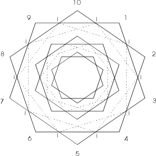
To do so, we must necessarily add a layer of complexity to our description. As previously mentioned, there are two additional subset double pentagons (decagons) which are intimately related to the M and m sets, but are offset by a 180 rotation. To refer to all 5 decagon sets (that is 10 pentagons), we will label them as follow:
| (LARGE) | (MEDIUM) | (SMALL) |
| MAJOR M | MAJOR m | MAJOR µ |
| MM | Mm | Mµ |
| spoken as major-major | spoken as major-mid | spoken as major-minor |
These are the same set described earlier...note that they all are major.
The two new subset, minor decagons are:
| (SUBSET LARGE) | (SUBSET MEDIUM) |
| minor M | minor m |
| µM | µm |
| spoken as minor-major | spoken as minor-mid |
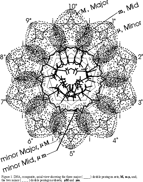
| The axial view of the double helix reveals:
Zone 1. a radial outer zone Zone 2. a clearer mid zone Zone 3. a complex inner zone |
zone lA Figure. 2. |
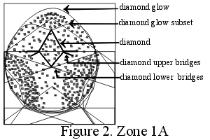 |
| The outer radial zone 1 consists of:
lA. 10 diamond shapes 10 diamond glow areas 10 upper and lower diamond connecting bridges In addition,each diamond glow has 2 (subsets) smaller glows, top left and right. |
||
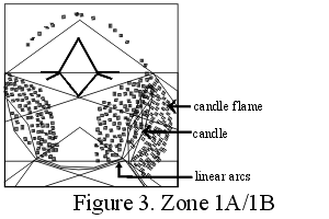
| lB. (interspersed between lA)
10 candle shapes 10 candle flame forms ( which are interconnected with inner, linear arcs to give a semi-circular pattern below (inward) in zone 2 |
zone lB _____________ zone 2B Figure. 3. |
 |
| The middle clearer zone 2 consist of:
2A. a clear space between the lower portion of the diamond shape 2B. 10 sets of linear arcs interconnecting the lower portions of the candle shapes and simultaneously forming a linear, undulation of pentagon and hexagon tops circling the center, the 20 heads of which match the 20 subset diamond glows in zone lA with the radial, linear elements and the second clear zone just above zone 3. |
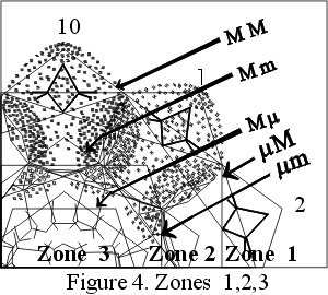
zone 1,2 & 3 Figure 4. |
 |
| The inner, complex zone 3 consists of:
entirely of lines and spaces, partially mimicking and resonating with the pattern of the middle zone 2, but with a great deal more complexity...do to the large number of pentagonal and hexagonal forms at the center. |
Combining the visual and pentagonal views (Figure 1.) reveals:
Zone 1, the outer radial zone is contained in the decagon sets MM & µM
Zone 2, the middle clear zone is contained in the decagon sets Mm & µm
Zone 3, the inner complex zone is contained in the decagon set of Mµ .
To draw Zone 1:
-Draw the MM, interconnect ipi to give µM
-1A (diamonds) are located at hourly positions on a 10 hour clock by drawing Mm and µm decagons and extending the ipi of this set, locating the lower side of each diamond. The upper side of each is found by rotating the fulcrum from the lower side of the top diagonal of the other diamond.
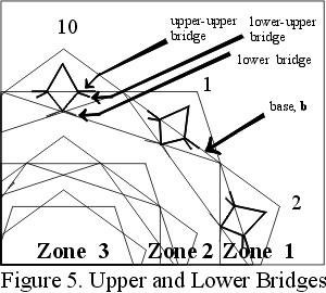
-The upper-upper diamond bridge goes from the diamond at hour 1 to the tip of diamond at hour 2.
-The lower-upper diamond bridge goes from the lower-upper diamond at hour 1 to the lower-upper diamond at hour 3 (which exactly follows the base, b, of MM if it is rotated 18°° ), Figure 5.;
-The lower bridge is simultaneously defined by the same lines of the lower-upper bridge;
-The diamond glow is defined by the MM, as is the subset, Figure 5.;.
Zone 1:
This represents the phosphate group (PO4) of the sugar-phosphate backbone, which defines the helix. The foundation of the sugar-phosphate backbone is the sugar (deoxyribose, a ribose sugar with two oxygens). A pentagonal molecule, interconnected by the phosphate groups running on it's outside and interconnected to its complementary helical strand on the inside by the nucleotide bases.
The combined unit of one phosphate, one sugar and one base is called a nucleotide. The nucleotides are spaced at 10 per 360° turn of the helix. Each complementary strand (which runs in the opposite direction), thus reveals a double pentagon pattern of 10 nucleotides per every 360° rotation and the dual helixes are justified so the clear decagon pattern is reinforced in the MM and µM sets.
The pentagon-based deoxyribose sugar defines the decagon, including the pentagonal pattern of the cross bridging of the diamond patterns of phosphate groups. The deoxyribose sugar is not itself flat and planar with the axial view, but rather is orientated more vertically (perpendicular to the page) as see in the more standard side view of the double helix.
The 10 candle shapes and flame forms represent the 10 sugars of each helical strand per one full rotation. They are located at the intersection of the µM and µm sets. The upper limits of the candle flame are defined by the tips of the µM set, the lower limits by the base of the µm set, while the candle runs in the center of its flame between these two points, its angular sides are formed from lines linking the intersections ( 1/2 hours) at 4 hour intervals. The angular lines represent the pentagonal sugars as seen on edge.
Zone 2 and Zone 3:
-The outer points (hours) of the Mm decagon defines the major void area;
-The same points on the µm decagon spot the lower crossings of the candle;
-The line forming the base of the same pentagon defines the border of the base pairs;
-The base of the Mµ pentagon defines the inner ring.
Actually, the middle and inner zone combined represent the paired bases inter-connecting the double helical strands. The four bases come in two forms:
a. Purines: have a double ring structure (pentagon & hexagon), Figure. 6.;
b.Pyrimidines: have a single ring structure (hexagon), Figure 6.
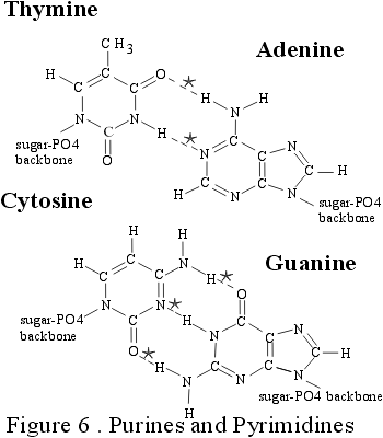
*connects three dimensionally to specific complementary bases via hydrogen bonds here.
The Mm and µm sets define the ring structure of the base which is attached to the deoxyribose sugar (for purines it is a pentagon, for pyrimidines it is a hexagon). The inner linear zone Mµ defines the second ring structure (for purines a hexagon) and/or the side chains and the hydrogen bonds connecting the opposing bases. As seen, the bases lie perfectly flat and planar to the cross sectional view down the axis of the double helix molecule. To note is that there is always a purine base matched with a pyrimidine base and thus there is always a pentagonal ring structure (contributed by the purine base) in every base pair set, and as such, therefore, 10 such times around the clock, i.e. one for each hour. Base pairs always match as A:T & G:C.
The ability to recognize, incorporate and replicate oneself...especially amidst the sea of diversity, the winds of chance, the lands of opportunity...is to organize one's energies to overcome those forces of entropy. The DNA double helix molecule accomplishes such feats by means of the very same self-recognizing, self-incorporating and self-replicating attributes of the very geometry upon which it is built. The DNA Master Chart has given us a glimpse, and I am sure it is only a partial view, on how that might be done. The beauty of all life forms is a reflection of the inner beauty of that natural organizing logic and form within.
REFERENCE
This work is based on simple concepts on elementary geometry, driven by a love for pattern seen in numbers, as in Nature. The simplicity defies the diversity, yet it is through a combination of fixed, almost rigid and constant symmetry with limited, asymmetric variables that the fabric of life is woven. From the original computer generated imagery of DNA by the pioneering Robert Langridge (http://www.cgl.ucsf.edu/home/rl/index.html) over 20 years ago, I analyzed and subsequently deconstructed the image pattern into a number of geometric forms, the primary ones being a series of concentric double pentagons. In 1995 I rebuilt the pattern on the surface of a large sculpture ("Out of Africa, from mime to five, DNA").That same year I had started an ambitious paper: "The Geometry of Music, Art and Structure...linking Science, Art and Esthetics". Revised in 1996 and further revised and condensed in the final published version in 1998. My work on the Geometry of DNA was a small part of this work. I more fully developed GoDNA in the next interim work: "Pattern in Number...from Primes to DNA" in 1997, and have since readied it for formal publication in Part 2 of "The Geometry of Music, Art and Structure...linking Science, Art and Esthetics". The ideas presented in the white papers were part of my gallery shows at the former Montage Gallery in Portland, Oregon in the mid 1990's.
"The Geometry of Music, Art and Structure...linking the esthetics of Art and Science", Part 1,
was presented at the 8th ICECGDG (International Conference on Engineering Design Graphics and
Descriptive Geometry, sponsored by the International Society for Geometry and Graphics, the Japan
Society for Graphic Science, and the Engineering Design Graphics Division of the American Society
for Engineering Education; hosted by the College of Engineering, The University of Texas at Austin), 1998.
Published in the Proceedings of the 8th ICECGDG, Vol. 2, pg 378, ISGG, Tokyo, Japan, 1998
Sloane, NH and York,JL, Review of Biochemistry, The Macmillan Co, Toronto, 1969.
Copyright© 2001-06 Reginald Brooks, BROOKS DESIGN. All Rights Reserved.
click to go to Special Graphics Section: digital 2D works: GoDNA